When faced with serious medical conditions in infants, neonatologists often turn to point-of-care ultrasound (POCUS) as a life-saving tool. POCUS offers targeted, bedside scans that produce rapid results, unlike traditional ultrasounds that provide comprehensive diagnostic reports, which must be interpreted by a radiologist.
Technicians train to perform medical procedures like POCUS with a device called a training phantom, or a realistic simulation of anatomy or tissue that allows trainees to practice without risk to patients.
Through a year-long collaboration with Johns Hopkins Hospital, fourth-year students Allison Booher (biomedical engineering) and Alexa VelezFonseca (chemical and biomolecular engineering) and third-year students Grace Guan (applied mathematics and statistics) and Sebastian Tabares Erices (mechanical engineering) observed that most training phantoms do not adequately prepare technicians to perform ultrasounds on infants.
The student team was matched with project partners at JH Hospital in the two-semester course Multidisciplinary Engineering Design, where they worked closely with Dr. May Chen, director of the Neonatal Point-of-Care Ultrasound Program. The team also interviewed technicians and observed POCUS training sessions at the JHMI Children’s Center, through which they learned that the size and accuracy of the training phantoms were inadequate to train technicians on many neonatal procedures.
“Training phantoms for POCUS are typically adult-sized and do not reflect the unique anatomy of neonates,” said Guan. “Commercial, neonate-sized training phantoms usually only focus on one function and are extremely expensive or employ simplified designs that do not entirely replicate the look of various anatomical features.”
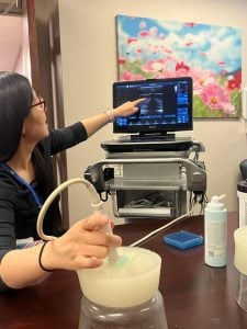
The team observes a point-of-care ultrasound on a training phantom.
To address issues of accuracy and affordability, the team designed a neonatal training phantom for POCUS that is anatomically accurate, accessible, and inexpensive.
The team’s first challenge was to scale down the anatomy of a training phantom, while also maintaining detail and complexity. “A baby’s heart is about the size of a walnut and the veins are around 3 millimeters in diameter and collapse under very little pressure,” Tabares Erices said.
Aside from nailing down the finer details of infant anatomy, materials and manufacturing processes created additional challenges. To successfully train technicians to perform POCUS, the phantom needed to resemble human tissue “to an ultrasound, not just to the human eye,” Tabares Erices said. “Using the wrong material or even minor manufacturing artifacts, like tiny bubbles or string, can create shadows and white spots that make it hard to identify anatomy with the ultrasound.”
Through experimenting with various processes, including 3D printing, molding and casting, working with polymeric silicone, and studying POCUS techniques, the team developed a multi-step method to produce various molds and outer shells that mimic actual tissue and affect echogenicity, or the ability of materials to reflect ultrasound waves. This process “was driven by frequently working and testing with end users for feedback and rapid incorporation and interation,” said VelezFonseca.
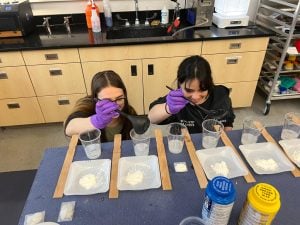
The team experimenting with materials in the iteration process of designing their neonatal POCUS training phantom.
While they were able to develop a detailed and responsive prototype, multiple casting steps made the process complex and expensive. The team then considered how to manufacture their neonatal training phantoms more efficiently and accessibly, including providing documentation and video tutorials of the process to clinicians so that they can reproduce phantoms on their own. “Our end goal is for any clinician to be able to manufacture these high-fidelity models themselves without breaking the bank,” Tabares Erices said.
The students are now designing a line of phantoms, including a neonatal pericardial effusion model, a neonatal vein model, and a neonatal spinal model, all of which are novel for their size or improve upon existing commercial phantoms.
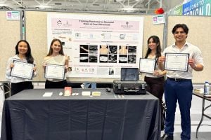
The team recently presented their prototype at Design Day on Tuesday, April 28 and were awarded the Dean’s Design Award for the Center for Leadership Education. The award recognizes undergraduate projects that identify and solve complex engineering problems, and demonstrate creativity in the design process, effective communication skills, and a thorough understanding of the underlying engineering principles.