Researchers have developed a method for turning commonly used steroid drugs into tiny particles that release their medicine slowly and predictably, a step that could reduce dosing frequency and side effects for many conditions.
The results appear in Science Advances.
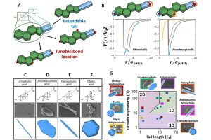
Steroids are molecules derived from the metabolism of cholesterol in the body. They are characterized by a steroid core and a tail containing the side group. Corticosteroids, a class of synthetic steroid, serve as anti-inflammatory therapeutics for treating conditions such as arthritis, asthma, multiple sclerosis, and COVID-19. However, their effectiveness often comes with significant drawbacks. Patients frequently require high doses or repeated treatments, leading to unwanted side effects such as weight gain, osteoporosis, eye problems, and increased infection risk. In addition, frequent drug dosing is a major contributor to patient noncompliance. This new study aims to address these issues by redesigning how these drugs are delivered in the body.
“Our goal was to design a system that reduces how often patients need treatment while maintaining strong therapeutic effects,” says Thi Vo, assistant professor in the Department of Chemical and Biomolecular Engineering.
The team worked with bile acids and corticosteroids—two types of steroid-based molecules. Using a modified chemical process, they created uniform microparticles that gradually release the drug as they break down. In laboratory and animal tests, these particles showed consistent performance and maintained their biological activity.
A key discovery was understanding the process of how the particles form. The researchers found that small changes in the chemical “tail” of steroid molecules, specifically how easily it gains or loses a proton, are crucial for particle formation.
“Ionization of the side group of molecules is essential for particle formation,” says Vo. “Without it, the molecules do not assemble into particles.”
They also discovered that hydrogen bonding—a weak attraction between molecules—helps hold the particles together. By adjusting the length and structure of the molecules’ side groups, the team was able to control the shape and size of the particles, forming rods, sheets, or spheres.
In experiments, the particles were tested for their ability to break down fat cells. In lab-grown human cells, the particles caused fat cell destruction in a dose-dependent way. In mice, injections of the particles led to a significant reduction in fat tissue without major side effects. “All particle types caused a significant reduction of fat pad mass compared to controls,” says Vo.
Importantly, the tests found that the particles worked well with various additives, such as gold nanoparticles, gold ions, or the common anti-inflammatory compound salicylic acid. This flexibility suggests that the method could be adapted and scaled for different drugs. The team also demonstrated that drugs that previously could not form particles could be modified to do so, opening the door to broader applications.
The findings could have significant implications for future treatments. By enabling drugs to release slowly and predictably, this technology could reduce dosing frequency, limit side effects, and improve patient compliance. It may also allow scientists to design drug particles with specific shapes and behaviors tailored to different diseases.
“These results enable us to not only predict what molecules can form into particles but also design and optimize particle morphology before fabrication,” says Vo. “We hope that the research points to a future where drug delivery is not only more efficient but also more personalized.”
The team for this research includes University of Michigan’s Ann Arbor’s Oluwaseun D. Akanbi, Michael L. Felder, Daniel Kupor, Lisa J. Bain, Crystal Sanchez, and Omola Eniola-Adefeso; University of Nebraska Medical Center’s Jiachen Feng, Luana Janaína de Campos, and Martin Conda-Sheridan; and University of Delaware’s Hanieh Safari.
Johns Hopkins University’s Thi Vo provided data curation, formal analysis, funding acquisition, investigation, methodology, project administration, resources, software, supervision, validation, visualization, and writing.
This research was financially supported by The National Science Foundation, the National Institutes of Health, Fast Forward Medical Innovation Mi-Kickstart and mid-stage MTRAC grants, the University of Michigan Cellular Biotechnology Training Grant, and startup funding provided by Johns Hopkins University. Simulation performed for this research used resources of the Advanced Research Computing Facility (ARCH) at Johns Hopkins University.