Chemotherapy is a powerful weapon against cancer, but certain cells resist treatment by entering a dormant stage called senescence. These therapy-induced senescent (TIS) cells may become resistant to therapy and even turn aggressive and spread. Detecting TIS cells early could be pivotal in preventing their growth, but current screening methods fall short in speed and accuracy.
New advanced microscopy techniques developed by a team of researchers at Johns Hopkins University and Politecnico di Milano, Fondazione Istituto Nazionale dei Tumori, and Consiglio Nazionale delle Ricerche in Italy may change that, paving the way for clinicians to identify these cells early and adjust treatment options. The team’s results appear in Science Advances: “Noninvasive morpho-molecular imaging reveals early therapy-induced senescence in human cancer cells.”
“Our work demonstrates the potential to transform anticancer treatment research,” said Ishan Barman, an associate professor of mechanical engineering at Johns Hopkins Whiting School of Engineering. “Integrating these microscopy methods could help clinicians make more informed, timely treatment decisions.”
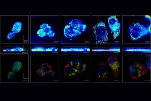
The team combined three cutting-edge, label-free microscopy techniques—coherent Raman scattering, multi-photon absorption, and optical diffraction tomography—to observe and analyze TIS cells in their natural environment. Unlike conventional identification methods, these advanced tools enabled the researchers to observe and study the cells’ shape, structure, and physical and chemical characteristics throughout their lifecycle.
“It’s important to note that we conducted these observations without the use of invasive labels, ensuring that the cells’ natural states were preserved,” said Dario Polli, an associate professor of physics at Politecnico di Milano.

Ishan Barman
These techniques revealed significant changes that occur in the TIS cells. For example, in 24 hours, the cells’ mitochondria, the “powerhouses” that produce energy, rearranged themselves. Within 72 hours, the cells also began overproducing fatty molecules called lipids and flattened and grew larger. This analysis allowed the team to establish a clear timeline for the development of these changes.
The researchers say that their new rapid-live screening microscopy techniques hold enormous promise for the future of cancer studies.
“While further research is needed, our results suggest label-free microscopy could become a vital tool to boost treatment efficacy and prevent tumor recurrence by detecting TIS cells early. This would allow clinicians to adjust therapeutic strategies before drug-induced senescence enables cancer to develop resistance,” said Italia Bongarzone, a senior research assistant at Fondazione Istituto Nazionale dei Tumori.