Farhad Pashakhanloo, PhD ’16, a postdoctoral fellow in the Institute for Computational Medicine, enjoys using his camera to document life as it happens.
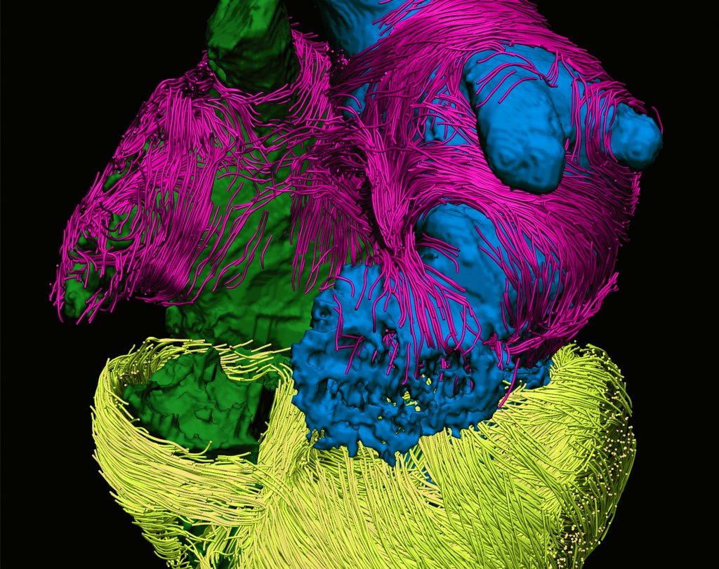
His most widely known image yet, however, wasn’t captured with his Nikon D610. Instead, with the help of a clinical MRI scanner at the National Institutes of Health, Pashakhanloo spent more than 60 hours creating an image of a human heart, which has been chosen to grace the cover of Nature Reviews Cardiology every month for the next year. (For variety and aesthetic appeal, each cover image will be in a different color scheme.)
“I obtained this image using what is called diffusion tensor MRI as part of my doctoral work and decided to enter it into a contest being held by the journal,” says Pashakhanloo, who works in the laboratory of Natalia Trayanova, the Murray B. Sachs Professor in the Department of Biomedical Engineering. “I thought it was visually very interesting, and the journal editors obviously agreed.”
Even more interesting, however, is the accomplishment the image represents: the first time, ever, that a human heart’s atrial chambers—the two upper chambers, which receive blood from the body and pump it to the lower chambers, the ventricles—have been imaged in their entirety to reveal the orientation and arrangement of bundles of heart cells.
“This is important because our data will be used to create accurate computational models of patients’ hearts, which we can then use to better understand sources of rhythm abnormalities, such as atrial fibrillation,” says Pashakhanloo, who completed his PhD in biomedical engineering last summer. He was advised by Trayanova and Elliot McVeigh, formerly the BME chair and now at the University of California San Diego.


