Fighting Cancer with Shiny Nanocrystals
Quantum dots, nanometer-sized semiconductor crystals that glow brightly in pure, intense colors, hold promise for many medical applications. They are brighter and longer lasting than the organic dyes used today to image tumors, and they hold promise for treating tumors via light-triggered heating.

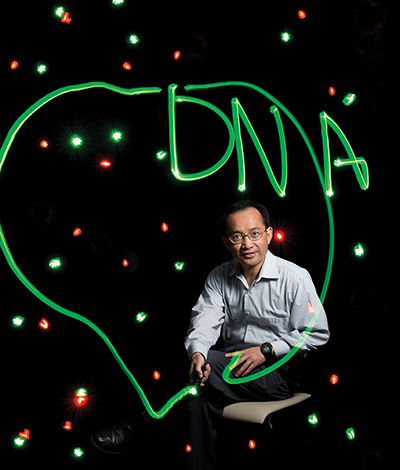
The nanocrystals could also be harnessed to help doctors catch cancer early. Jeff Wang, associate professor of mechanical engineering, is developing a technique to do just that. One place doctors can look for early warning signs of cancer is DNA, the helical strand carrying the unique genetic blueprint of every individual.
Long before cancer symptoms become apparent, invisible signals called genetic biomarkers appear on DNA. They can be an inherited or mutated gene sequence or a chemical change at a specific location on DNA.
But spotting cancer biomarkers is not easy. They are present in body fluids such as blood and sputum in extremely small concentrations. Wang is working on an ultra-sensitive quantum-dot-based technique that would allow doctors to detect the presence and quantity of cancer genetic markers in body fluids. A larger quantity means higher cancer risk.
The technique could lead to personalized cancer medicine. “By identifying the markers, doctors could provide treatment plans tailored to a patient’s genetic information,” Wang says. Another important use for the test would be in monitoring whether a therapy is working effectively.
Wang says the nanocrystals should help spot genetic biomarkers in body fluids with more speed and precision than the polymerase chain reaction (PCR) method commonly used today.
PCR involves making billions of copies of a piece of DNA in order to spot target gene sequences. “Most of the time, standard PCR isn’t sensitive enough to look at DNA circulating in blood and present in other body fluids,” he says. “You have to perform the test multiple times to detect the gene sequence, and the method is inconsistent.”
Wang has been working on a quantumdot- based cancer detection test for three years with colleagues at the Johns Hopkins Kimmel Cancer Center. They introduce a protein and a fluorescent dye into the fluid sample along with quantum dots. The molecules attach to the biomarker-carrying DNA, and pin it to a quantum dot. Each nanocrystal can carry tens of dye-tagged DNA strands.
When the researchers shine blue laser light on the fluid, quantum dots transfer the light to the dye molecules, which start shining. Analyzing the fluorescence signal reveals how much biomarker is present.
The test has proved its mettle in detecting a cancer marker called DNA methylation. Methylation stops the release of tumor-suppressor proteins, making it easier for cancer cells to multiply. Early findings show that the test could detect 50 or fewer target DNA strands in sputum samples from lung cancer patients.
Wang is now perfecting the technique using real-world fluid samples taken at the Kimmel Cancer Center.
More recently, he has developed a quantum-dot-based tool that could help doctors monitor whether a drug is fighting cancer as expected. The test relies on spotting changes in the mobility of the nanocrystals in fluid.
He has found a way to precisely “tune” the mobility of quantum dots depending on the number of target DNA strands tethered to the dot. Measuring the mobility indicates the amount of biomarker DNA present in fluid.
“This should give doctors a way to measure minute biomarker quantity changes,” Wang says. “If a drug isn’t working, there won’t be a decrease in biomarker level.”


