


Light defines our world. It lets us see, gives us warmth and energy, and allows us to grow food.
Electromagnetic radiation. Photons. Packets of energy. Waves. Particles. There are few facets of our modern lives that light, in its many forms, does not touch. Light enables the integrated circuit chips in our computers, the displays on our smartphones, and the ultrahigh-speed Internet we rely on. Indeed, light-based technologies have revolutionized everything— from health care and manufacturing to defense and communications.
Advances in optics (which deals with light’s behavior and properties) and photonics (which has more to do with light’s applications) have become “central to modern life,” according to the National Research Council, which recently issued a report calling for a National Photonic Initiative to foster collaboration between industry, government, and academia to identify and advance key areas of photonics research.
Whiting School engineers are part of the national thrust to further the understanding and application of optics and photonics. By pushing the frontiers of light, they are pioneering techniques that could advance cancer diagnosis, surgical tools, hydrogen fuel production, and microscopic imaging. And that’s just a start.
Light-Based Surgical Tools
Thousands of disease sufferers, accident victims, and war veterans gain new lives every year thanks to organ transplants, reconstructive surgery, and the reattachment of body parts. These tricky operations require surgeons to sew together blood vessels that are a millimeter or less in width, and a few tens of microns thick. Not only are the vessels tiny, they are semitransparent when blood is removed.
Surgeons performing such delicate suturing procedures must use microscopes to see that minuscule depth and then move their needle through it. But microscopes have their limitations because they do not offer adequate depth perception.
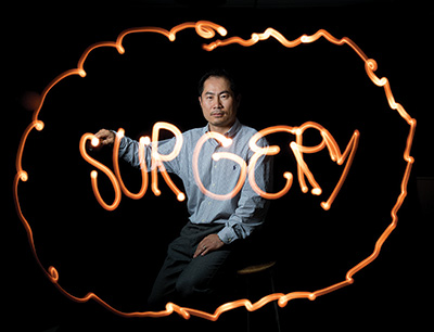
“A common mistake that surgeons make is to go through both walls of the vessel,” says Jin Kang, chair of the Department of Electrical and Computer Engineering, and the Jacob Suter Jammer Professor of Electrical Engineering. “So instead of suturing two parts of a vessel to each other, they end up sealing the vessel.”
A fiber optics expert, Kang is using laser light to measure submillimeter distances and to create highly accurate, detailed images of tissue features just microns in size. He is also perfecting a surgical tool using this light-based sensing system. “It’s very similar to ultrasound but we’re using a laser beam instead of sound waves,” Kang says. “While ultrasound has a resolution of hundreds of microns, our system has a resolution of a few microns.”
In any imaging technology, the smallest detail you can resolve depends on the size of the wavelength used to make the image. Light, with wavelengths less than a micron, can create images with submicron resolution.
Kang uses low-power light from a nearinfrared laser with wavelengths ranging from 700 nanometers to 1300 nanometers. An optical fiber delivers the laser light to the imaging site and also collects the light reflected from different parts of the tissue. A computer processes the collected light signals to recreate an image.
Instead of peering through the eyepiece of a microscope, a surgeon suturing blood vessels could place the optical fiber sensor near the surgery site and look at an image on a screen to guide her needle. Kang’s collaborators at Wilmer Eye Institute recently tested the imaging system during a delicate surgery of a rabbit’s eye, taking pictures of the different parts of the animal’s retina.
Kang and his team eventually want to extend their fiber-optic imaging system for surgeries deep inside body tissue. Right now, the system can only create images of features present up to two millimeters below tissue surface. Strong light scattering by body tissue keeps light from penetrating beyond that distance.
He also has other plans in the works. “We want to use faster lasers and detectors to achieve a real-time, higher-resolution 3-D imaging system,” he says.
Unlike the big machines used for today’s imaging techniques, the laser and fiber-optic approach entails a simple, inexpensive, portable tool. “The lasers and sensors are in a backend system that is the size of a shoebox,” Kang says. “The optical fiber, meanwhile, is a fine thread 100 micrometers in diameter. The nice thing is, the optical fiber could be integrated into any surgical tool.”
In fact, he has already incorporated his optical fiber sensor into a “smart” surgical tool in collaboration with Hopkins’ Engineering Research Center for Computer-Integrated Surgical Systems and Technology. The tool is designed to compensate for the near-invisible tremors of a surgeon’s hands and keep the surgical instrument steady.
The end of the optical fiber is mounted on the tip of a surgical instrument and is connected to a motor that controls the axial movement of the instrument tip. The system gauges the tip’s position relative to tissue and then adjusts it. Those tiny adjustments go unnoticed by the surgeon, who can use it to perform delicate surgical procedures more safely and precisely.
Others are developing similar 3-D surgery-assisting devices, but those are more complicated, Kang points out. They involve external sensors that are not embedded on a surgical tool. The sensors track the motion of the surgical tool tip and then do 3-D position control.
The technology behind Kang’s tool has been licensed to robotic surgery companies such as Intuitive Surgical. It could find use in surgeries on the sensitive neural circuits of the brain; the fine structures of the inner ear; and the retina, the thin light-sensitive tissue lining the back of the eye, Kang says.