


Q: How will engineering advance our understanding of metastasis, and ultimately lead to improved treatments and therapies for cancer
Metastasis, or the spread of cancer, starts when a cancer cell detaches from a tumor, enters the bloodstream, and then invades tissue elsewhere in the body to start the site of a secondary tumor. These secondary sites are responsible for the majority of cancer-related deaths. If we can understand the critical steps in metastasis and better understand all the processes involved, we have the opportunity to halt the spread of cancer by blocking just one of the steps.
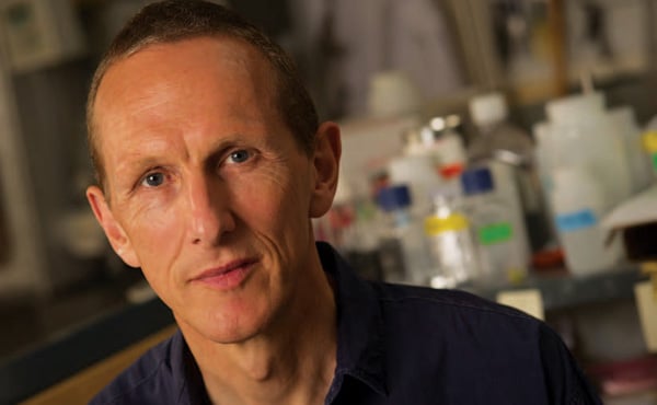
”In my lab, we have created the first lab-on-a-chip aimed at understanding metastasis. It’s about the size of a smartphone, and it re-creates the entire microenvironment of a tumor and blood vessels necessary to study the spread of cancer.” Peter Searson
One of the greatest challenges we face when studying metastasis is the difficulty of tracking individual cancer cells as they move from a primary tumor to a secondary site. To address this difficulty, we are engineering every component involved in the process— the tumor, the surrounding tissue, and the blood vessels—in a microdevice that mimics a vessel and surrounding tissue.
In my lab, we have created the first lab-on-a-chip aimed at understanding metastasis. It’s about the size of a smartphone, and it recreates the entire microenvironment of a tumor and blood vessels necessary to study the spread of cancer. The device contains a hydrogel material, representing the extracellular matrix, into which we have formed a cylindrical channel about the width of a human hair. This provides the template for a blood vessel. Endothelial cells then are plated to the surface of the cylinder, forming an artificial blood vessel. Each end of the vessel is connected to a flow system, allowing perfusion.
Using this device, we can place a ball of cancer cells in the vicinity of the artificial vessel or inject individual cancer cells into the extracellular matrix. Because the device is transparent, we can observe the cell migration into the vessels in real time. By perfusing molecules into the device, we alter the microenvironment in ways that simulate what happens during each stage of metastasis. For example, we can change the concentration of collagen in the gel and make the matrix stiffer or softer. Or, we can change the type of tumor cells or introduce other molecules, drugs, and substances and see how these variables affect the results. This knowledge may provide researchers with new methods to halt the spread of cancer.