


 A brain imaging technique developed by engineering researchers at Johns Hopkins’ Center for Imaging Science has revealed subtle but important differences in the brains of autistic children.
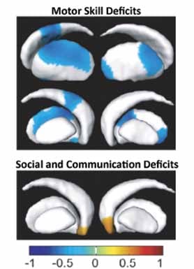
Children with the disorder had small shape differences in a group of brain structures called the basal ganglia, a part of the brain associated with motor skills and learning. The differences were associated with problems involving motor control, performance and imitation of learned motor skills (for example, combing hair and brushing teeth), and social and communication deficits.
Michael I. Miller, the Herschel and Ruth Seder Chair in Biomedical Engineering and director of the Center for Imaging Science, used a technique he developed with his colleagues called large deformation diffeomorphic metric mapping (LDDMM). LDDMM uses standard magnetic resonance images to show in detail where structures are in the brain. The technique allows computers to look not just for overall size differences in brain structures but for subtle differences in shape as well.
“We basically try to understand the brain and its parts. You’d like to be able to make a quantitative statement about how a typical brain looks, and then in what way a diseased brain is atypical and therefore quite different,” Miller says.
For the autism study, images of the basal ganglia of 32 children with autism spectrum disorders were compared with those of 45 children without the disorder. The MRIs of each child’s brain were divided into 100 million voxels—cubes 1 millimeter on a side—and the voxels were analyzed by an IBM supercomputer at the Whiting School’s Institute for Computational Medicine.
The study found three separate structural differences between autistic children and other children, and each difference was associated with a specific deficit. In children with autism, poor motor control was associated with shape change in one part of the basal ganglia, difficulty with learned and imitative behavior with another, and poor social communications skills in yet another.
“These findings actually make sense in terms of what we understand about basal ganglia function,” says Stewart H. Mostofsky, associate professor of neurology at the School of Medicine, who co-authored the paper. The deformations occur in parts of the basal ganglia that are known to play a role in the specific aspects of motor control and social communication that are impaired in these children, he says.
The work contributes to a basic understanding of autism spectrum disorders, which affect an estimated 500,000 children in the United States. Such knowledge, Mostofsky says, might one day help doctors individualize therapies according to a patient’s brain structure.