


Gabor Fichtinger’s robot-assisted device offers a less-invasive way to detect and treat prostate disease.
Using a simple robotic arm tipped with a needle, Gabor Fichtinger and his research team are hunting a killer that accounts for nearly 31,000 deaths a year in the United States. The disease is prostate cancer, the country’s third leading cause of mortality among men.
Currently, early detection of this cancer involves a manual clinical procedure called a transrectal ultrasound (TRUS) guided needle biopsy. However, the male prostate gland, about the size of a walnut, has a delicate suspension system that can easily be damaged if a biopsy needle exerts too much squeezing or pushing, Fichtinger explains.
Despite its wide use, TRUS-guided biopsy has been shown to miss the presence of cancer in approximately 25 percent of cases. Given that approximately 65 million prostate biopsies are performed annually in the United States, revealing around 220,000 new cases a year, this 25 percent rate of failure is unacceptable to Fichtinger.
A Johns Hopkins University associate research professor, Fichtinger has joint appointments in the Whiting School of Engineering departments of Computer Science and Mechanical Engineering, and in Radiology at the School of Medicine. He also is the director of engineering at Hopkins’ Engineering Research Center for Computer-Integrated Surgical Systems and Technologies (ERC CISST), where he leads a team researching computer-assisted systems for inserting thin delivery devices though the skin to target locations.
“Using TRUS, if we are dealing with a cancer that’s the size of a sugar cube, there’s no guarantee that we can see it,” Fichtinger says. “And even if we can see it, the deformation and dislocation caused by using a handheld device gives us no assurance that we will hit the prostate accurately.”
“This is a robot that doesn’t even work like a robot, but still does a procedure that nobody had been able to do before.” Gabor Fichtinger
A Sharper Focus for Diagnosis
Fichtinger earned his PhD in computer science in his native Hungary, at the University of Budapest. Drawing from his early background in computer graphics and biomedical visualization systems, as well as his engineering expertise, Fichtinger began devising an alternative to TRUS after arriving at Hopkins. His goal was to have a sharper eye—and a steadier hand—to make prostate disease diagnosis and research more effective. Robotic assistance could provide more precise and predictable movement and needle placement. But Fichtinger also wanted the system to provide enhanced imaging to enable a higher rate of disease detection. To do so, he needed a robotic device that could operate inside of a conventional closed MRI scanner.
The challenge of working with an MRI scanner was threefold, according to Fichtinger. First, such scanners do not allow access to the patient during imaging. That meant his device had to work within the extremely cramped space of the MRI’s core, where conventional medical robots and mechanical linkages cannot. Second, due to the MRI’s magnetic field, which is 300,000 times more intense than that of Earth, any ferromagnetic materials or electronic devices could heat up and become hazardous to the patient. And third, a real-time in-scanner guidance method was needed to operate the device. “Therefore, our primary objective was to develop a prostate biopsy system that coupled superior imaging quality with accurate delivery hardware, inside a conventional MRI scanner,” says Fichtinger.
“It was really an amazing, precise procedure…”—Bob Siblo

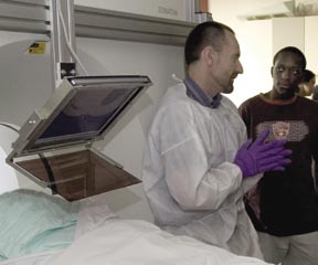
Bob Siblo never expected to encounter a robot. Then again, he never expected to have cancer. But when a screening test in 2004 showed that he had high levels of PSA (prostate specific antigen), indicating the likelihood of prostate cancer, Siblo began looking at treatment options. At the National Institutes of Health in Bethesda, Maryland, he decided to become a test subject for a novel procedure, one that employed MRI scanning and targeted radiation therapy using the Johns Hopkins In-MRI Prostate Robot.
“My entire treatment process only lasted from March to July,” recalls Siblo. “It was really an amazing, precise procedure that made me feel very comfortable. I never felt any discomfort or side effects.”
Since his treatment, Siblo, who lives in Annandale, Virginia, has nothing but good news to report. “In just one year, my PSA levels have gone from 9.1 down to 2.1,” he says. “So everything at this point seems to be fine.”
Sophisticated Solution, Made Simple

Just two years after its conception, the prototype was ready. The In-MRI Prostate Robot is elegant in its simplicity yet remarkably sophisticated in its design. “That’s the beauty of it,” Fichtinger says with a laugh. “This is a robot that doesn’t even work like a robot, but still does a procedure that nobody had been able to do before. Although a lot of thinking went into its development, it is as simple as it needs to be to do the job.”
Using actuation cables, the physician or technician controls the robot. Once its extendable arm is inserted rectally into the patient, the robot uses real-time MRI-based position sensors to calculate precisely the sequence of movements needed to bring its needle to an intended target point.
The robot’s general-purpose needle delivery system not only can do biopsies but can also deliver capsules. These capsules could place markers to help target subsequent therapies, or deliver a genetically engineered virus or a radiation therapy seed.
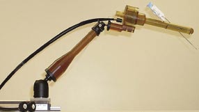
The MRI scanner can constantly collect high-fidelity images and sends them immediately to the robot’s treatment monitoring computer. The resulting 3D representation of the device is superimposed on the anatomic images. This enables the physician to monitor and adjust the motion of the device toward its target.
Beyond its diagnostic and therapeutic uses, the new robot offers a significant application as a research vehicle, Fichtinger says. “We now can do long-term follow-ups with patients to take measurements, or do biopsies to track any pathological changes. With an MRI, there are also endless possibilities for functional imaging,” he notes. “We can look at a number of indicators related to the prostate that we would have no way to see in ultrasound.”
Rapid Development with the Right Team
Spurred by a grant from the National Institutes of Health, the nowpatented In-MRI Prostate Robot moved from concept to human trials in a remarkably short period—just 24 months. Fichtinger credits this rapid development to one critical factor. “This project could not have been done without the collaboration of other specialists in a wide range of disciplines,” he says. Among the principal contributors he cites Louis L. Whitcomb, professor of Mechanical Engineering; Ergin Atalar, professor of Radiology and of Biomedical Engineering; Axel Krieger, a PhD candidate in Mechanical Engineering; and Robert C. Susil ’97, an MD/PhD student in Biomedical Engineering. “It’s definitely been a team accomplishment,” says Fichtinger.
To learn more about the ERC CISST, visit engineering.jhu.edu/~erc-cisst and to learn more about Gabor Fichtinger’s other projects, visit www.cisst.org/~gabor.