
Medical imaging techniques, such as echocardiogram, CT, and MRI, have greatly improved the diagnosis of anatomic and functional heart abnormalities. Though these methods are effective, they are too time consuming and expensive for use in screening large numbers of people. Thus, there is an urgent need for an efficient and accurate screening method, which will be used complementarily to the medical imaging techniques.
The StethoVest
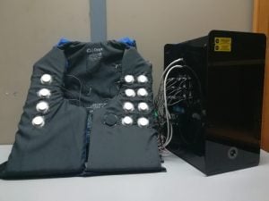
Christos Sapsanis, a doctoral candidate in the Department of Electrical and Computer Engineering, is part of a team working to devise a method of accurately and inexpensively screening people for cardiovascular issues that can be detected through heart sounds. The group, based in the lab of Andreas Andreou, a professor of electrical and computer engineering, has developed the “StethoVest,” a prototype wearable phonocardiographic (PCG) system designed to record sounds emitted from the heart from multiple areas of the chest simultaneously.
The StethoVest uses a lifejacket with sensor nodes placed on the inside lining, which are connected to a readout system. The nodes then record the patient’s heart sounds in various regions of the chest around the heart. The eventual plan is for the full readout system, and all the electronics, to be embedded within the vest and for the results to be sent to the patient’s phone, making it portable, wireless and user friendly.
“The vision of the StethoVest is for the stethoscope to transform into a wearable sensor node and phone in an interconnecting point for live listening, saving the data online and displaying the diagnosis automatically,” Sapsanis said. “The final goal is to provide a complete system from heart sound acquisition to an accurate and fully automated diagnosis, minimizing the need of a physician in the screening part and expanding the target market to the general public.”
The StethoVest is in the early stages of being used at Johns Hopkins Hospital, where one of the key features has been its ability to register single recordings that can be conducted in multiple body postures, such as standing, sitting, squatting, or lying down.
Andreou’s group is collaborating on the project alongside William R. Thompson, a pediatric cardiologist and associate professor at the Johns Hopkins School of Medicine, as well as Rajat Mittal, a professor of mechanical engineering, whose lab focuses on computational fluid dynamics. Sapsanis says that the ease with which these collaborations have come about has been crucial for the development of the StethoVest.
“In addition to giving us access to patients’ database, Dr. Thompson has given me valuable insight into this field, while Dr. Mittal’s group has helped us understand the effect of valvular abnormalities on the blood flow and sound generation from a computational perspective,” Sapsanis said. “Both of these contributions have been essential. In the Andreou Lab, we try to identify a problem and then approach it with multidisciplinary efforts.”
If you would like more information on the StethoVest, check out the academic paper presented in IEEE Biomedical Circuits and Systems Conference in 2018.