
When Yufan He entered the Best Young Scientist Award competition at MICCAI: International Conference on Medical Image Computing and Computer Assisted Intervention, he knew the odds of winning were slim.
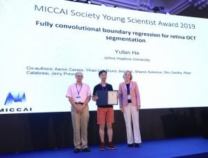
Yufan He on stage after being announced as a MICCAI Society Young Scientist Award winner
More than 500 papers were competing for the coveted award, which recognizes the highest quality papers that are first-authored by young scientists at the conference, which took place in mid-October in Shenzhen, China.
So, when He, a doctoral candidate in the Department of Electrical and Computer Engineering, learned that his paper—“Fully convolutional boundary regression for retina OCT segmentation”—was one of five winners, he was thrilled. That victory was even sweeter as He and Zongwei Zhou of Arizona State University were the only winners representing American institutions.
“It was really unexpected and exciting,” He said. “I didn’t expect that my work and presentation would be that highly recognized by the award committee. To me, winning this award means that I have a very good understanding of a problem and have proposed a novel method to solve it.”
He’s work focuses on Optical Coherent Tomography (OCT) image segmentation, which is an imaging method used to take cross-section pictures of the eye’s retina. Despite being a well-explored area, the practice has great limitations. To overcome these, He developed an improved method by using deep learning in a novel way.
“To quantitatively analyze medical images, for example, for acquiring retinal layer thicknesses, we need to segment each retinal layer from the image,” He said. “After segmentation, we can obtain statistics about the changes of each retinal layer and better understand the disease or the treatment.”
He’s method could have a major impact in the diagnosis of multiple sclerosis (MS). If a patient takes an OCT scan of their retina, his algorithm can produce quantitative results of their retinal thickness or lesion volume within a minute after finishing the scanning. From there, the clinicians can take the quantitative results and compare with the longitudinal data to better diagnose or monitor the disease.
For big data analysis, like segment tens of thousands images in the database, a fast segmentation algorithm is needed. As a result, He’s system has been used to analyze thousands of scans from people with MS in a short time.
“Our group has been working on OCT image analysis for people with MS for many years and I just continued working on this topic,” He said. “The deep learning method is an emerging technique that has many benefits in image analysis but it has disadvantages in giving structured output. I’m interested in overcoming the disadvantages of this method while maintaining its benefits for OCT image analysis.”
He chose to earn his doctorate at Johns Hopkins in order to work with Jerry Prince, the William B. Kouwenhoven Professor in electrical and computer engineering, who is also the director of the Image Analysis and Communications Lab. The advisor-advisee relationship has blossomed into a fantastic pairing, He says, and he is excited to see where it takes him.
“I love the research environment at Johns Hopkins,” He said. “Dr. Prince and Johns Hopkins encourage us to think both hard and independently. They’ve provided a great research environment for me to think deeply and work on something novel.”
Click the video below to watch He accept his award.