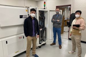
The Department of Materials Science and Engineering is the new home of the Advanced X-ray Imaging of Materials (AXIOM) system. The first of its kind, AXIOM provides two advanced capabilities for studying the structure of materials: X-ray phase-contrast imaging and high-energy x-ray diffraction microscopy. Although XPCI and HEDM have been available to researchers using synchrotron x-ray light sources at national laboratories, time on those facilities is limited. With the installation of AXIOM in the laboratory of Todd Hufnagel, Hopkins researchers will have unparalleled access to these advanced capabilities year-round.
Traditional x-ray imaging, or radiography, make use of differences in x-ray intensity resulting from differences in absorption to study the internal structure of materials. When examining a broken bone, for example, the dense bone absorbs x-rays more strongly than the soft tissue that surrounds it. X-ray phase contrast imaging (XPCI) extends this by making use of spatially-coherent x-ray source to observe differences in the phase of the x-rays, as well as their intensity. XPCI makes it possible to observe internal defects such as cracks and pores, even in materials that are weakly absorbing and provide poor contrast in traditional radiography. Prof. Hufnagel and Ryan Hurley from the Johns Hopkins Department of Mechanical Engineering plan to use AXIOM to study deformation and fracture of sand, sandstone, and other geological materials as part of the Materials Science in Extreme Environments University Research Alliance (MSEE URA), funded by the Defense Threat Reduction Agency (DTRA).
High-energy x-ray diffraction microscopy (HEDM) is a revolutionary new technique that makes it possible to look inside metals and other structural materials to develop a 3D “crystallographic map” of the grain structure. With HEDM the position, size, and crystallographic orientation of every grain in a bulk specimen can be measured. Because it is non-destructive, HEDM is idea for in situ studies of how the grain structure evolves during deformation. Hufnagel’s group will use HEDM to study fatigue failure in aluminum and other light-weight structural alloys.
The advanced features of AXIOM are enabled by its unique x-ray source. In a traditional x-ray source, high-energy electrons are accelerated towards a metal anode, which produces x-rays. The brightness of the x-rays produced is limited by the need to dissipate excess heat, which otherwise would melt the anode. To avoid this problem, the MetalJet source in AXIOM uses as its anode a thin stream of a liquid indium-gallium-tin metallic alloy. Because the anode is already molten, a tremendously bright ray spot can be produced by focusing an intense electron beam into a small spot on the liquid stream. Capabable of producing high energy x-rays (up to 160,000 electron volts) at high power (700 watts), the new source provides higher brightness for the XPCI and HEDM experiments than any other source outside of a synchrotron.
“We’re excited to do some really challenging experiments and to tackle complex materials problems with a more sophisticated way of looking at materials as they are changing,” says Hufnagel. “And we’ll be doing it right here at Johns Hopkins University.”