How are certain experiences, like enjoying a concert or watching a movie, reflected in neural activity across the whole brain? How do cancer cells in a tumor respond differently to therapies? Answering these questions requires technology that can track the dynamics of all cells of interest across the entire organ or organism.
For decades, researchers have worked to map and understand the complex activity patterns within tissues and organs, but they have relied on technologies that can only take static snapshots or monitor dynamics of a small subset of cells.
This is a challenge that Dingchang Lin, core researcher at the Institute for NanoBioTechnology and Assistant Professor of Materials Science and Engineering, set out to solve.
“In neurobiology research, the ideal scenario is to map as many neurons as possible across the whole brain, ideally every single neuron, but this is just nearly impossible using the existing tools,” Lin says., “It is challenging to interface with every single neuron at the same time, and it would be almost impossible to process that massive amount of data generated in real time.”
Now, published in Nature, Lin and his team at Johns Hopkins University have gotten much closer to this ultimate aspiration. The paper is “Genetically encoded assembly recorder temporally resolves cellular history.”
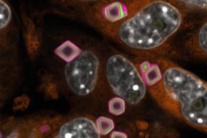
They approached this ambition by developing a genetically encoded intracellular recording platform, Granularly Expanding Memory for Intracellular Narrative Integration, or GEMINI. GEMINI turns a computationally designed protein assembly into an intracellular memory device to record individual cells’ activity histories. When expressed in live cells, GEMINI assembles predictably with minimal interference to cellular functions. As GEMINI particles grow, they capture cellular activities as fluorescent, ring patterns in the scaffolds, enabling imaging-based retrospective readouts. As the technology is genetically encodable, it can be deployed at a large scale across an intact organ.
“Just like tree rings that document climate changes from centuries ago, GEMINI creates ring-like molecular records of cellular histories inside cells,” says Lin. “By looking at GEMINI’s cross sections under a microscope, we can read what individual cells have experienced via these small, fluorescent ‘rings.’” In practice, GEMINI is an analog recorder that can quantify how strongly cell activity occurs, providing a measure of signal intensity across cellular processes. By integrating reliable timestamps, GEMINI enables chronological reconstruction of cellular histories and records when specific events happen inside the cell, down to a resolution of tens of minutes. It can also pinpoint certain phases of cell dynamics with roughly 15-minute resolution.
“Imagine a memory device that can record information in such details within every individual neuron in the brain,” Lin says. “Isn’t it cool that we make cells produce the entire recorder machinery themselves to document their own activity?”
In a mouse model of cancer, the team turned GEMINI on within cancer cells throughout an entire implanted tumor. As the tumor grew, GEMINI captured each cell’s activity history, while the tumors continued to grow normally.
“This means scientists can finally look back and monitor cancer cells across regions of the same tumor over time, rather than relying on the end-point snapshot,” said PhD student Jiaxi Lu, the co-first author of this study.
In a mouse brain, GEMINI documented changes in neural activity without disrupting the neuronal physiology or normal brain function. The animals behaved normally in tests of movement, coordination, and memory. This means that GEMINI could record the history of a brain seizure to be temporally resolved.
“GEMINI’s robust image-based readout makes it seamlessly integratable with various of fluorescence microscopy tools and protocols to reveal hidden details in physiological, pathological, developmental models and beyond,” said Yuqing (Eugene) Yan, a Kavli Distinguished Doctoral Fellow and the first author of this study.
The team sees two major areas of impact for this technology. First, GEMINI could advance fundamental research in cell biology and neurobiology by preserving spatiotemporal information across as many cells as one could imagine, providing opportunities to take a microscopic look at cellular dynamics in healthy and diseased tissues. Second, GEMINI might transform drug discovery by revealing, with unprecedented detail, how therapies affect individual cells within diseased organs or tumors.
So far, GEMINI has been implemented across an entire tumor xenograft and locally in the mouse brain. Next steps include scaling the platform to the whole brain and enabling efficient, accurate readout of the recorded information. In this effort, advanced microscopy technology and AI-based decoding tools will play key roles.
“In the future, we are excited to further explore the potential of the GEMINI system to reveal the temporal and spatial dynamics of tumor progression, metastasis, and adaptation to diverse microenvironments,” Lu says. “Tumor biology remains an area of intense research interest, and we are excited about uncovering the mechanisms underlying tumor heterogeneity. We believe that the GEMINI system could serve as a powerful complementary approach alongside existing methodologies to help elucidate temporal, spatial, and activity-based heterogeneity in tumors.”