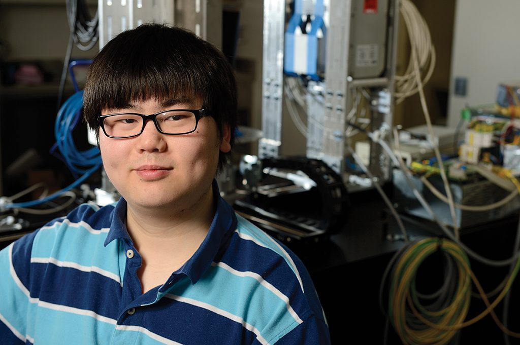
 Minutes before his doctoral oral exam, Qian Cao checked his email and got a thrilling surprise: The PhD candidate had been chosen for a coveted Howard Hughes Medical Institute International Student Research Fellowship, awarded to just 45 international predoctoral students studying in the United States.
“I was overloaded with happiness that day,” remembers Cao, now in his third year in the Department of Biomedical Engineering.
The award of $43,000 annually through the fifth year of his graduate program will help fund his work to improve the technology used for CT scans to give this imaging method the potential to view minute bone changes that signal the start of osteoarthritis.
Osteoarthritis, Cao explains, is growing steadily more prevalent in the United States as the population ages. Some estimates suggest that more than 67 million individuals will have osteoarthritis by 2030.
However, no diagnostic test is available that can catch the disease in its early stages, when interventions to treat this condition or slow its progression could be most effective. Preclinical research suggests that tiny changes in trabecular bone—spongy tissue found at the ends of the body’s long bones and in the pelvis, ribs, skull, and vertebrae—might signal the onset of osteoarthritis. Yet CT’s current resolution limit of approximately .5 millimeters prevents researchers from confirming this hypothesis and eventually using trabecular bone changes for early diagnosis.
To improve CT technology, Cao’s research has focused on development of a new generation of CT systems that will achieve improved resolution, thanks to the implementation of X-ray detectors based on complementary metal-oxide-semiconductors. “They’re the Ferraris of X-ray detectors,” Cao says, “offering both higher resolution and lower noise.”
Along with higher resolution comes an incredible onslaught of data, which necessitates new algorithms to interpret this information. That’s why Cao and his colleagues are also working on developing new software and analysis methods.
Cao’s research is under way in the I-STAR Laboratory at Johns Hopkins, where he works with mentor Wojciech Zbijewski and Jeffrey Siewerdsen, professor of biomedical engineering, on the development of new 3-D medical imaging methods. “Qian’s work will help break the conventional limits of spatial resolution in CT,” says Siewerdsen.


