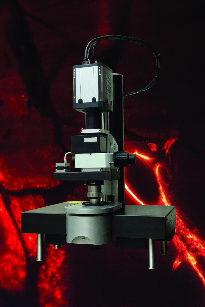
A new 3-D microscope has arrived on the Homewood campus. The cutting-edge tool condenses what had previously required months of work into just hours, and it allows researchers unprecedented views of organs, tissue, and even live specimens.
The selective plane fluorescence light sheet microscope is one of the first operational on the East Coast and the only one in Maryland. Purchased with a grant from the National Institutes of Health, it cost $360,000.
It illuminates specimens from the side, shooting two perfectly aligned planes of light across an object, illuminating a wafer-thin slice of the whole while the camera captures an image—thousands of times over as the specimen moves through the light. The result: A 3-D image of the full object.
Audrey Branch, a fellow at the Kavli Neuroscience Discovery Institute (co-directed by BME Director Michael I. Miller, MS ’78, PhD ’83), was one of the first to use it. She says she cried when she saw the first pictures of a mouse brain, its individual neurons glowing red, and its spindly dendrites, too—showing quite clearly the links between those cells. “It feels so amazing to see the brain in a way that no one has ever seen it before,” she says.
Web Extra
Watch Branch use the microscope.


