


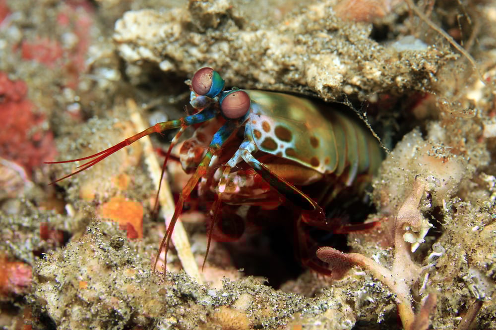
The mantis shrimp’s bulbous eyes are about as complex and curious as they come. Tiny, tubelike nanostructures allow the shrimp to sense colors and other properties of light that are invisible to human eyes.
Viktor Gruev, MS ’00, PhD ’04, has emulated the mantis shrimp’s visual system to design image sensors for early cancer detection. He has made a simple, compact prototype camera that could allow doctors to spot precancerous tissue that would be easily missed before the cells become visible tumors.
When light bounces off objects, some of it is always polarized. Polarized sunglasses filter out this part to get rid of glare. “But instead of throwing it away, we want to capture and quantify it,” says Gruev, an associate professor of electrical engineering at the University of Illinois. That’s because the polarization and colors of light carry information about the objects the light bounces off.
Cells in healthy tissue, for instance, have an organized structure that scatters light very differently from diseased tissue, in which cells are disorganized. “Their color and polarization signature are different from each other,” he says.
Scientists have, in the past, made imaging devices that sense polarization. But these devices have been bulky, costly, and complicated. So Gruev worked with marine biologists and sensory neurobiologists to understand how the tubelike nanostructures that decorate mantis shrimp eyes filter and sense polarized light. Then, he and his team created nanowires in the lab that emulate those structures. By integrating the nanowire polarization filters in front of complementary metal-oxide-semiconductor and charge-couples device detectors found in conventional cellphone cameras, the researchers made compact polarized image sensors.
The sensors are small enough to fit at the tip of an endoscope. The researchers have tested the camera on mice with colon cancer. Today, doctors use endoscopes to look for suspicious tissue and then do a biopsy. But the tissue or polyp has to be a certain shape to be visible. “In some patients, a flat tissue region develops into full-blown cancer polyps in a short span of time,” Gruev says. With the new tool, the researchers were able to spot those flat, precancerous lesions that look normal to the human eye.
Collaborating with colleagues at Washington University in St. Louis, Gruev has incorporated the camera into high-tech goggles that let surgeons see the exact boundaries of tumors. This helps them to precisely remove cancer cells and spare healthy ones, making surgeries safer and reducing repeat surgeries. Pilot tests during breast cancer and melanoma surgeries have proved that the wearable technology highlights tumors with 100 percent sensitivity. Gruev is now planning larger studies and hopes to improve the goggles so that they can detect microscopic lesions.