


On the human cerebral cortex, sulcal and gyral curves snake across the surface in a Hydra-like array of sizes and configurations. Could mathematics be used to match those curves and fully automate the brain mapping procedure? To recognize patterns in an image with ambiguous regions, in which order should tests be performed, and when should testing stop? These are the kinds of questions that intrigue researchers in the Center for Imaging Science, which delves into the realm where mathematics, computer science, biomedical engineering, and electrical engineering converge.
Advanced geometry, statistics and probability theory, functional analysis, harmonic analysis, optimization theory…these are a few of the branches of advanced mathematics enlisted in imaging science. This emerging field is at the forefront of research in connection with developing medical, military, and security-related software, to name just a few applications. It’s all about getting information of a semantic or diagnostic nature in an automated way. Security personnel could use image interpretation to scan a scene or a photogaph to find a face in a crowd. It would be useful in analyzing MRI or other images to help doctors in their diagnoses of disease. The process involves comparing shapes from the images and, in medical analysis, determining normal versus pathological.
CIS’s diverse team of faculty members work together in Clark Hall to solve some of the most complex problems associated with image interpretation. In terms of mathematics, the research involves interpretation of shape, and it’s a complicated problem. “The first thing you need to do in image interpretation is define what shape means, mathematically,” says Carey Priebe, professor of Applied Mathematics and Statistics. “But, that’s still unknown territory.” That’s because shape involves infinite-dimensional parameters, which makes statistical analysis literally impossible. But it doesn’t mean the problems are impossible to solve or at least approach solving.
CIS, established in 1998 as a Whiting School of Engineering research center, is part of the Whitaker Biomedical Engineering Institute. Michael I. Miller, a highly cited researcher in computational anatomy, is the Center’s director as well as professor of Electrical and Computer Engineering, and the inaugural Herschel L. Seder Professor of Biomedical Engineering. (He was profiled in “High Performers” in the Fall 2002 Johns Hopkins Engineer). In one project at CIS, Miller and colleagues are investigating image interpretation in the relatively new field of brain mapping.
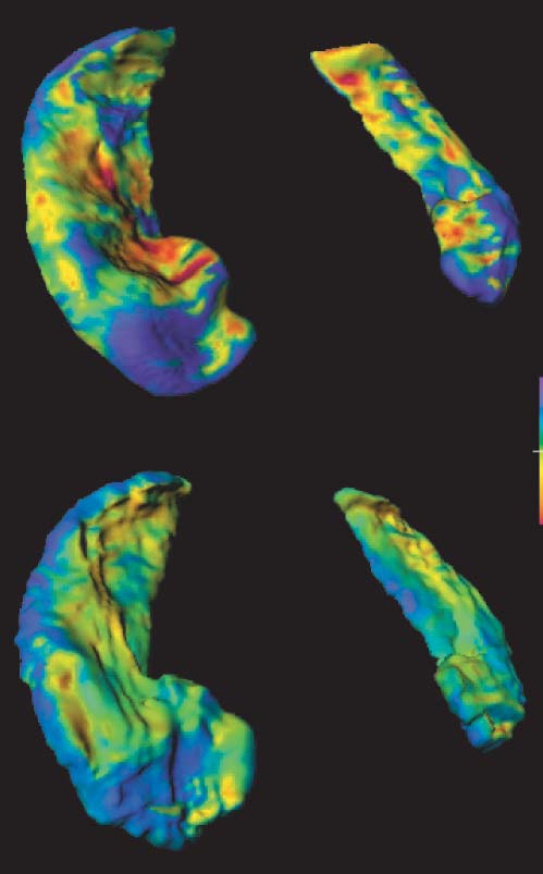
Says Donald Geman, professor in the Department of Applied Mathematics and Statistics, “The single biggest problem we’re working on is computational anatomy, the analysis of organic shapes, with applications in neuropsychiatry and other fields.” The team is exploring the connection between shape and deformity of the hippocampus region in the brain, to eventually enable doctors to detect patients with DAT (dementia of the Alzheimer type) or Alzheimer’s disease.
To do this, CIS is developing software using a mathematical technique called “deformable templates,” complex algorithms that enable the comparison of shapes and that analyze normal versus pathological. There’s still a long way to go in developing this technique, along with finding a better understanding, and eventually, a mathematical theory that defines shape in the same way that we now have for motion.
One of the newest members of the CIS team, E. Laurent Younes, associate professor of Applied Mathematics and Statistics, joined Hopkins last summer from the Centre de Mathématiques et Leurs Applications (CMLA) at Ecole Normale Supérieure de Cachan in France. Younes will add his expertise to the study of shape deformation in brain research, as well as statistical analysis of natural images, developing a mathematical understanding of how such images are built.
And this January, CIS welcomed René Vidal, assistant professor of Biomedical Engineering. Vidal recently received his PhD in electrical engineering and computer sciences from the University of California, Berkeley. His research includes among other topics, segmentation of static and dynamic scenes, multiple view geometry, and omni-directional vision.
“The contributions of faculty from all of the core departments exceed my own wildest expectations,” Miller observes. The academic model being developed to build research teams that integrate multiple academic departments “uniquely positions CIS to make contributions to many of the large-scale scientific efforts now under way throughout the nation, including the Bioinformatics Research Network, TeraGrid computational efforts, and large-scale integrative biomedical computing efforts.”
When all is said and done, imaging science comes down to the mathematical analysis that enables image interpretation. “The real action, the unexplored territory” with image science, explains Geman, “is with semantic and diagnostic analysis. It’s a big issue and one that looks as though it won’t be solved anytime soon.”
For upcoming seminars and other information about the Center for Imaging Science, visit cis.jhu.edu