


Scientists have long known that the cell membrane—the thin layer surrounding each cell—plays a crucial role in cell activities and communications. What hasn’t been clear, though, is how various proteins that are part of this membrane can dynamically change their location and grouping on the cell surface in response to various signals.
Understanding this mechanism could provide a deeper comprehension of cellular dynamics in general and also pave the way for targeted medical interventions for diseases related to cell signaling and migration.
A team of Johns Hopkins researchers—collaborating with researchers from Osaka University and RIKEN Center for Biosystems Dynamics Research, Japan—discovered a new method for selectively sorting certain proteins into distinct regions of cell membranes, providing insights into how cellular components organize themselves on a large scale during different physiological activities.
The researchers introduced a mechanism called “dynamic partitioning,” which operates on the idea that specific proteins can naturally or in response to external signals gather or organize themselves on the cell membrane. This occurs by adjusting their movement in different areas of the membrane.
“This discovery helps us to finally understand how proteins end up in different parts of the cell membrane and dynamically readjust their location as and when required. Our results provide a new lens through which we can understand how signaling events are tightly controlled, as cells communicate, migrate, or divide,” says team leader Pablo A. Iglesias, the Edward J. Schaefer Professor of Electrical and Computer Engineering at the Whiting School and interim head of that department.
The team started the project after discovering that certain proteins, believed to be uniformly spread across the cell membrane, actually consistently undergo dynamic polarization during various cellular events. “We were intrigued by this surprising result and wanted to dig deeper,” says Tatsat Banerjee, PhD ’24, lead author of the study, which appeared in Nature Communications, and former PhD student in the Whiting School’s Department of Chemical and Biomolecular Engineering and the School of Medicine’s Department of Cell Biology.
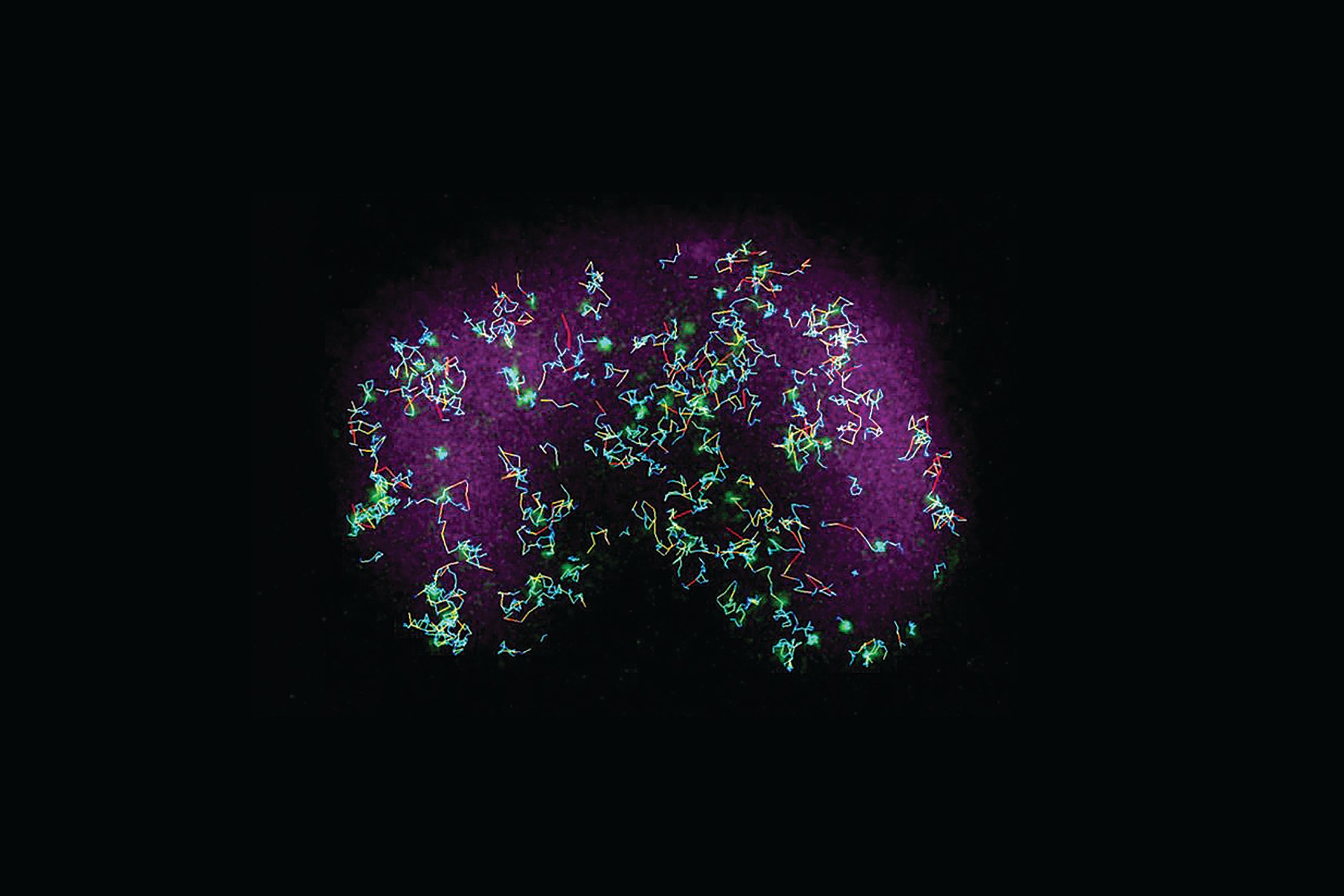
Banerjee and his team then used advanced microscopy techniques to observe and control events inside live Dictyostelium amoeba and mammalian immune cells in real time, with detailed resolution at the subcellular level. When they combined computer simulations and real-life experimental results, they found that many proteins in cells move selectively based on the organization of the membrane and how specific proteins sense and interact with its spatial variations.