


 When movie audiences in 1966 thrilled to watch a diminutive submarine race through a man’s arteries to save him from a deadly blood clot, The Fantastic Voyage was exactly that: an incredible story ripped from the pages of a science fiction novel.
Half a century later, the notion of deploying miniaturized tools into the body’s tiniest conduits and organs is no longer either fantastic or incredible: It’s happening.
“For more than 50 years, people have been talking about doing this, but we have actually done it.” David Gracias
One of the newest and most exciting examples of this comes out of the laboratory of David Gracias, the Russell Croft Faculty Scholar and an associate professor in the Department of Chemical and Biomolecular Engineering. A team led by Gracias, along with postdoctoral fellow Evin Gultepe and gastroenterologists Eun Shin and Florin Selaru of the Department of Medicine, has— for the first time ever—sent hundreds hundreds of untethered, dust-particle sized devices into the digestive system of a living animal to collect tissue for biopsies.
“For more than 50 years, people have been talking about doing this, but we have actually done it,” says Gracias, co-author on a paper about the research that appeared in recent issues of Gastroenterology and Advanced Materials.
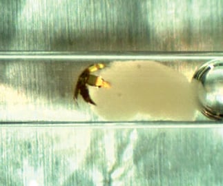
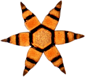
The ultrathin chromiumand- gold instruments called “mu-grippers,” each measuring less than a millimeter in diameter, can collect living cells from hardto- access places in the body without being attached to any wires or tubes, and without being powered by batteries or any external sources. Instead, Gracias and his team cleverly designed the grippers to respond to thermal and biochemical signals.
Stored on ice before the procedure, the mu-grippers’ six three-jointed digits extend straight out from the center “palm.” But once deployed via endoscope into the warm GI tract, the grippers’ polymer-coated joints soften, causing them to attach to mucosa and excise tissue samples. They are then collected by a magnet inserted through the endoscope, so the DNA can be harvested and examined for mutations and signs of cancer or other pathology.
Taking a biopsy this way is far superior to the conventional method, according to Selaru, who also is a molecular biologist.
“A typical cancer-surveillance endoscopic session for patients … involves taking about 30 random forceps samples of colon tissue, two at a time, which is time-consuming. The endoscopist can’t take more samples than that because it will cause undue trauma to the tissue. But with the mu-grippers, we can get literally hundreds of samples from various parts of the colon with little or no trauma,” he explains.
Even more important is the fact that the greater the number of tissue samples taken, the greater the chance (statistically) of finding mutations-in-the-making.
“We used a mathematical model that showed that it would take up to 1,000 samples to make sure you don’t miss a peanut-sized lesion,” says Gracias, who also is affiliated with the Institute for NanoBioTechnology. “When you consider that, it’s not surprising that when you sample only a small part of the colon, you might miss something vitally important.”
As promising as the technology is, though, the team has more work to do before it can be tried in humans.
“We have provisional patents on the design, and [the mu-grippers] are very easy and inexpensive to make,” Gracias says. “As we move toward this becoming a medical device, there is much finetuning to do. But in the end, we think this will be a real paradigm shift in the way biopsies are done, and that’s really exciting.”