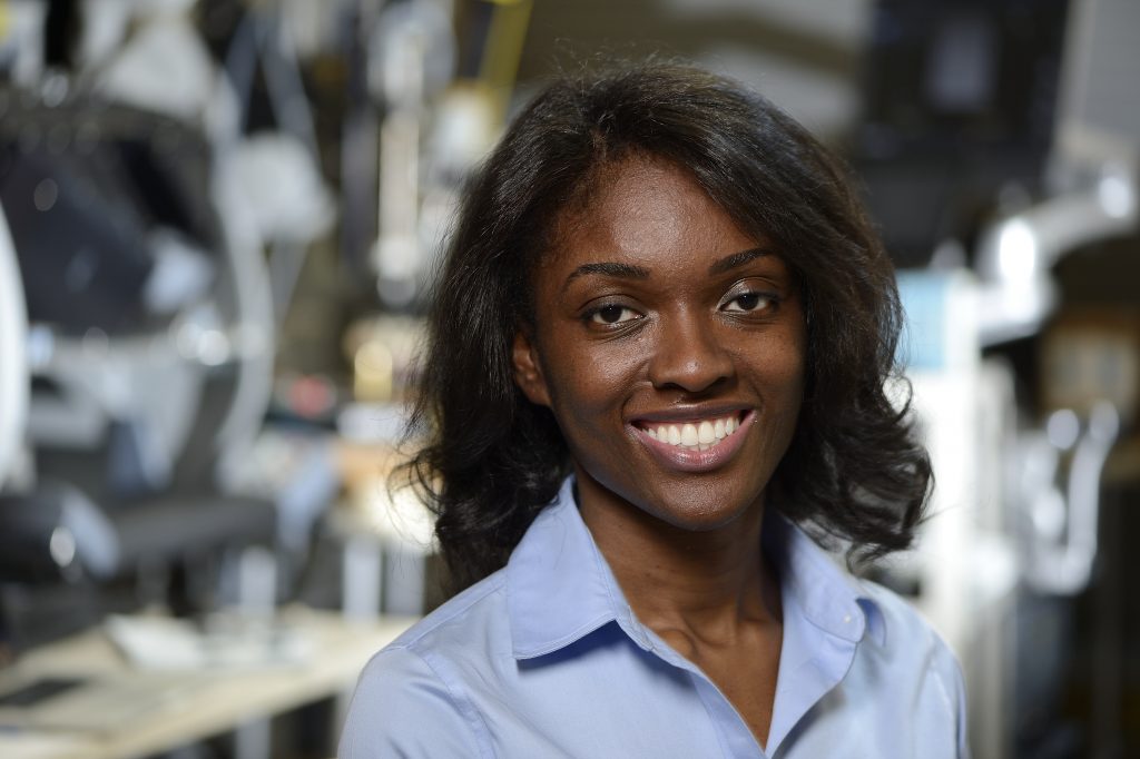
Minimally invasive surgeries are tricky. When bone or tissues obstruct the endoscopic camera’s vision, surgeons can accidentally injure blood vessels. Muyinatu A. Lediju Bell is designing a new image-guided surgical system that could give surgeons real-time visuals of the blood vessels, increasing precision and improving patient safety. For this and her other groundbreaking work on imaging, MIT Technology Review recently named Bell a 2016 Innovator Under 35 (see p. 4).
The system is based on photoacoustic imaging, an emerging technique that uses light and sound to produce medical images. It involves firing laser pulses into tissue, which expands and contracts from the small temperature rise, producing sound waves that are detected with ultrasound receivers. The signals are then processed to form images.
Bell, a newly appointed assistant professor of electrical and computer engineering, is integrating photoacoustic imaging into a da Vinci surgical system. This FDA-approved surgical system has three robotic arms that a surgeon controls from a console.
Bell has patented a novel signal-processing algorithm that produces high-contrast images, providing clearer pictures for real-time guidance of robotic surgery.
Traditional photoacoustic systems tend to generate cluttered images and need to have a fixed distance between the light source and acoustic receiver. Bell has patented a novel signal-processing algorithm that produces high-contrast images, providing clearer pictures for real-time guidance of robotic surgery. The new way to process sound signals also allows more flexibility between the light source and acoustic receiver. “For example, you can attach the light source to one arm of the da Vinci and the ultrasound probe to another arm,” she says.
Bell and her students in the Photoacoustic and Ultrasonic Systems Engineering Lab have done just that, through collaborations with the Laboratory for Computational Sensing and Robotics and with funding from the National Institutes of Health and the National Science Foundation Computational Sensing and Medical Robotics Research Experience for Undergraduates program.
Bell plans to test her customized photoacoustic system for pituitary tumor surgeries at the Johns Hopkins Hospital through her affiliation with the Carnegie Center for Surgical Innovation. The pea-sized pituitary gland sits behind the nose bridge and is flanked by arteries; minimally invasive surgery on it is done through the nose.
“We will insert the light source into the nose, keep the ultrasound probe on the patient’s temple, and see how well we can visualize the blood vessels located behind the bone,” she says.


