People
| Graduate Student | Zeng Zhang |
| Project Supervisor | Prof. Joseph Katz |
| Project Collaborator | Prof. Misun Hwang (Children’s Hospital of Philadelphia) |
Introduction
We aim to advance ultrasound localization microscopy (ULM) for high-resolution vascular imaging and quantitative blood-flow measurement. ULM reconstructs microvascular structures by localizing and tracking contrast microbubbles in CEUS images, enabling vascular mapping at spatial scales far below conventional ultrasound resolution. This capability is particularly important for studying cerebral microcirculation, where small changes in microvascular structure and flow can provide critical information about brain perfusion and hemodynamic function.
A major limitation of ULM is that each microbubble appears in the ultrasound image as a blurred trace that is much larger than the actual bubble diameter. Therefore, the accuracy of the final vascular map depends strongly on how precisely the bubble center can be identified from noisy and overlapping traces. Conventional approaches, such as blind deconvolution and other localization algorithms, can improve bubble detection, but their performance degrades when the point-spread function varies spatially, when background noise is high, or when neighboring bubbles and vessels are closely spaced.
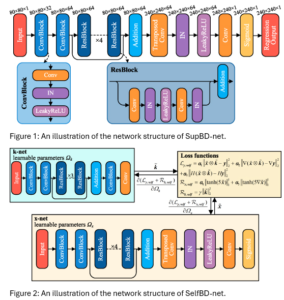
To overcome these limitations, we developed two residual-learning-based deep-learning frameworks: SupBD-net (fig 1), a supervised super-resolution blind deconvolution network, and SelfBD-net (fig 2), a self-supervised network designed to adapt to unknown bubble-trace morphologies. SupBD-net is optimized for high-accuracy bubble-center detection under realistic imaging conditions, while SelfBD-net improves generalizability by estimating the unknown point-spread function directly from raw CEUS images. Together, these methods improve the spatial resolution, robustness, and quantitative accuracy of ULM-based vascular imaging.
Results
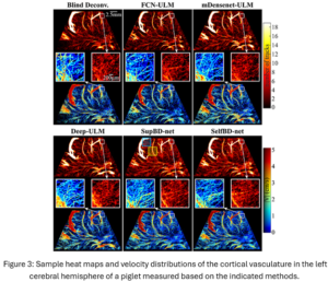
When applied to neonatal pig brain CEUS data, our methods generated high-resolution maps of cerebral microvasculature and blood-flow velocity. Compared with conventional processing, SupBD-net and SelfBD-net revealed finer vascular structures and better separated neighboring microvessels, including vessels spaced as closely as 0.15 wavelength in the cortical region. This demonstrates the practical value of the method for improving ULM-based cerebral vascular imaging.
The improved cerebral maps were enabled by more accurate microbubble localization. Our supervised method, SupBD-net, achieved the highest localization accuracy among the tested approaches, maintaining bubble-center errors below 0.1 wavelength even under challenging conditions with high noise, dense bubble concentrations, and large point-spread functions.
To improve robustness beyond the training conditions, we also developed SelfBD-net, a self-supervised framework that estimates unknown point-spread functions directly from raw CEUS images. Under unfamiliar bubble-trace morphologies where conventional supervised methods failed, SelfBD-net maintained localization errors below 0.15 wavelength, making it more generalizable across imaging settings.
Publication
Zhang, Zeng, Misun Hwang, Todd J. Kilbaugh, and Joseph Katz. “Improving Sub-Pixel Accuracy in Ultrasound Localization Microscopy Using Supervised and Self-Supervised Deep Learning.” Measurement Science and Technology 35, no. 4 (2024): 045701. https://doi.org/10.1088/1361-6501/ad1671.