
Subhrajit Das
PhD Student
Subhrajit is currently pursuing a Ph.D. in the Electrical and Computer Engineering (ECE) department of Johns Hopkins University under the interdisciplinary mentorship of two PIs, Prof. Ralph Etienne-Cummings, and Prof. Arvind Pathak. He received his M.S.E in Electrical and Computer Engineering in 2022 from Johns Hopkins University en route to his Ph.D. His research interest focuses on low-power, neuronal imaging devices, and applications of Deep Learning to elucidate key mechanisms underlying neuropathologies. He is currently developing an illumination control embedded circuit for low-power wireless multi-contrast mini microscopes to study neurovascular diseases in awake animals. Simultaneously, he is deploying deep learning models to study neurovascular diseases and their correlation with behavior.
S. Das, J. Senarathna, D. Yang, R. Chandra, A. Banerjee, and A. Pathak, Predicting animal behavior from multi-channel neuroimaging data, In ISNVD Annual Meeting on “Vascular Contributions of Healthy Aging and Dementia” Oral Presentation, 2022, New York, USA
S. Panda, C. Ganguly, S. Das, R. Mandal, and S. Chakrabarti, Performance of a leaky integrate-and-fire model vis-à-vis measured response of diseased neurons, In Proc. of IEEE International Conference on Advanced Networks and Telecommunications Systems (ANTS),2019, Goa, India.
C. Ganguly, J. Dasgupta, S. Das, S. Saha, T. Sarath, S. Panda, S. Chakrabarti, First Level Approximation of Measured Neuronal Response using a Leaky Integrate and Fire Model, In Proc. of. IEEE India Council International Conference (INDICON), 1-4, 2019, Gujrat, India.
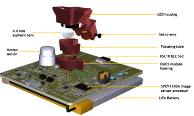
Evidence from preclinical and clinical experiments has shown that brain tumors alter the structure and function of the central nervous system during its progression and therapy. To elucidate the neuronal and hemodynamic changes accompanied by tumor progression, we need an imaging tool that can characterize these changes in vivo. We plan to build a ‘plug-n-play’ multichannel wireless microscope for microvessel resolution (~7-10 μm) in vivo imaging of a brain tumor for long hours. In the first phase, we will use an off-the-self eval board that uses the industry’s lowest BLE(RSL10) module to transfer images wirelessly and characterize the board before doing animal experiments. In the second phase, we plan to miniaturize this eval board keeping all the optics of the miniscope same.
ISNVD Travel Award (2022)
ISNVD 2022 Young Investigator Award finalists.